Specifications:
Product information: Medial Orbit
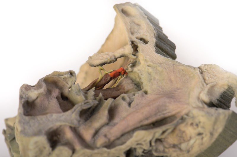
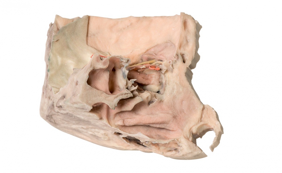
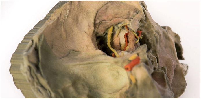
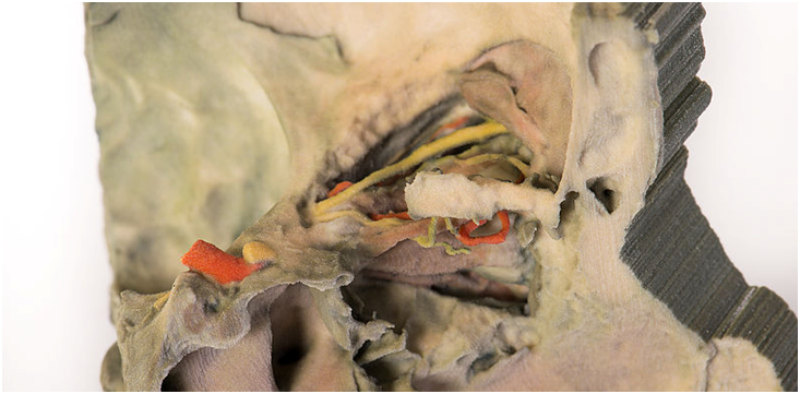
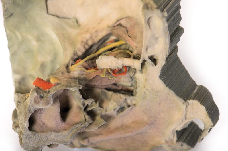
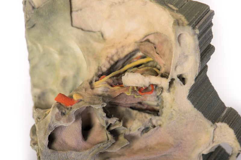
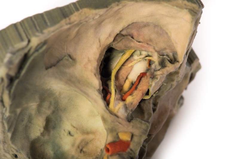
This 3D print displays the orbital contents and its close relations as viewed from the medial perspective when the majority of the lateral wall of the nasal cavity and the intervening ethmoidal sinuses have been removed. The posterior ethmoidal nerve (PEN) (a branch of the nasociliary nerve, CN V1) can be seen passing between the medial rectus (MR) inferiorly and the superior oblique muscle superiorly. A small piece of the orbital plate of the ethmoid bone (EB) has been retained to illustrate its path as it enters the posterior ethmoidal foramen. Other structures visible include the frontal nerve (FN), the sphenoid sinus (SS), the pituitary gland (PG) and the frontal sinus mucosal lining exposed after removal of the orbital plate of the frontal bone on the anterior roof of the orbit. The internal carotid and optic nerve are also visible within the cranium.
Catalogue: PDF
See more products OpenMedis brand.

Reviews
There are no reviews yet.