Specifications:
Further Information
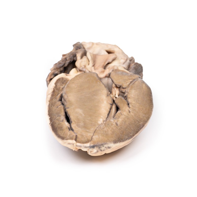
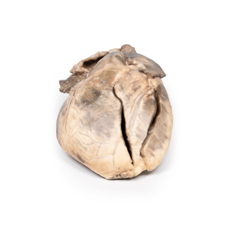
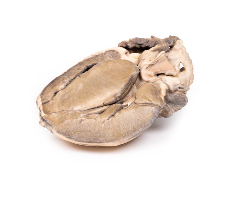
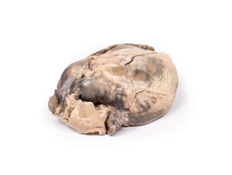
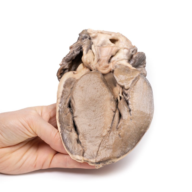
Subaortic stenosis is considered to be acquired rather than congenital and is suggested to result from an underlying defect in the architecture of the left ventricular outflow tract (LVOT). The defect may be such that the resulting turbulent blood flow leads to progressive thickening and fibrosis of the LVOT and the aortic valve. Progression of the disease will often lead to a hypertrophic cardiomyopathy secondary to the increased aortic pressure needed to be overcome by the left ventricle. Mild or moderate stenosis is often asymptomatic. As disease progresses and the stenosis becomes severe, symptoms such as exertional dyspnoea and syncope may become apparent. Investigation and subsequent diagnosis are often prompted by the presence of an ejection systolic murmur on physical examination. Echocardiography is used to confirm diagnosis. These days, definitive treatment of subaortic stenosis consists of surgical correction of the obstruction.
Catalogue: PDF
See more products OpenMedis brand.


Reviews
There are no reviews yet.