Specifications:
| Probes | Fundamental Frequency | 2.0MHz/2.3MHz/2.5MHz/3.0MHz/3.5MHz/4.0MHz/4.6MHz/5.0MHz/5.4MHz |
| Harmonic Frequency | 4.0MHz/4.6MHz/5.0MHz | |
| Fundamental Frequency | 4.0MHz/4.6MHz/5.0MHz/6.0MHz/7.0MHz/8.0MHz/9.2MHz/10.0MHz/12.0MHz/13.3MHz | |
| Harmonic Frequency | 8.0MHz/9.2MHz/10.0MHz, | |
| Fundamental Frequency | 3.0MHz/3.5MHz/4.0MHz/5.0MHz/5.4MHz/6.0MHz/7.0MHz/8.0MHz/10.0MHz | |
| Harmonic Frequency | 6.0MHz/7.0MHz/8.0MHz | |
| Fundamental Frequency | 3.0MHz/3.5MHz/4.0MHz/5.0MHz/5.4MHz/6.0MHz/7.0MHz/8.0MHz | |
| Harmonic Frequency | 6.0MHz/7.0MHz/8.0MHz | |
| Fundamental Frequency | 1.7MHz/1.9MHz/2.1MHz/2.5MHz/3.0MHz/3.4MHz/3.8MHz/4.2MHz/5.0MHz, | |
| Harmonic Frequency | 3.4MHz/3.8MHz/4.2MHz | |
| Fundamental Frequency | 2.0MHz/2.5MHz/3.0MHz/3.3MHz/3.7MHz/4.0MHz/5.0MHz/6.0MHz | |
| Harmonic Frequency | 4.0MHz/5.0MHz/6.0MHz | |
| 2D imaging mode | Gain | 0-100,Step 2 adjustable |
| TGC | 8 segment adjustable | |
| Maximum focus point | ≥7, which can be moved throughout the whole process. | |
| Speckle reduction | 0-5,5 level | |
| Space Synthesis | 0-2,2 level(Liner probe: 3 level, cardiac probe:0) | |
| Dynamic | 30-180,35 level,step 5 adjustable | |
| Line density | low、middle、high,3 level | |
| Frame correlation | 0-4,4 level | |
| Noise reduction | 0-5,5 level | |
| Edge Enhancement | 0-5,5 level | |
| Sound power | 2-10, 9 level | |
| Grey scale | 0-67, 67 level | |
| False color | 0-67,67 level | |
| Image style | Soft-Comparison,2 level | |
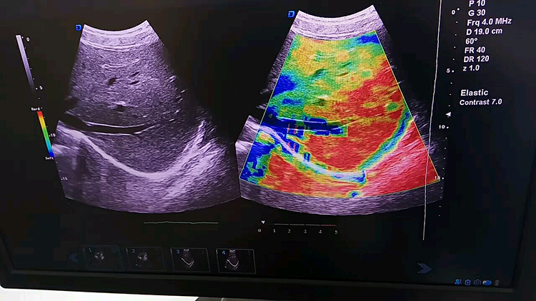
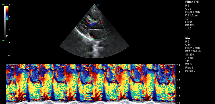
| Color Doppler imaging mode | Blood gain | 0-100,Step 2 |
| Parameter display | Velocity、Variance | |
| B-Restrain(B/W restrain) | 0-7, 7 level | |
| Speed Through | 0-8, 8 level | |
| Sampling number | 6-24, 7 level | |
| Blood flow preferred | 0-8, 8 level | |
| Filtering | 1-6, 6 level | |
| Sound power | 2-6, 4 level | |
| Noise reduction | 0-4, 4 level | |
| Smooth treatment | 0-4, 4 level | |
| Frame correlation | 0-6, 6 level | |
| Chromatography(Blood flow graph) | 0-37, 37 level | |
| Line density | Low-Middle-High, 3 level | |
| Frequency | 4 level adjustable | |
| Velocity | Minimum 0.4K,Maximum 40.5K | |
| Convex probe | 0.4K-4.3K-38.5K | |
| Linear probe | 0.4K-14.7K-39.0K | |
| Trans-vaginal probe | 0.4K-7.8K-39.7K | |
| Volume probe | 0.4K-4.2K-34.8K | |
| Micro-convex probe | 0.4K-10.3K-40.5K | |
| cardiac probe | 0.4K-7.8K-39.7K | |
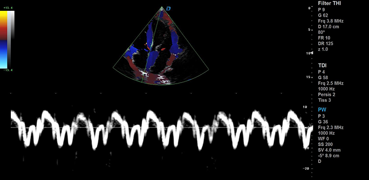
| Pulse wave Dopplermode | Gain | 0-100,Step 2 |
| Spectrum envelope function | real time automatic spectrum envelope, manual spectrum envelope, and other modes. The system automatically analyses and displays various data such as PSV, EDV, RI, PI, S/D, ACC, HR and so on. Can wake up or close | |
| Sample volume | 0.5mm~30mm | |
| Blood angel | -75—75 degree,Step 5 | |
| False color | 0-67, 67 level | |
| Dynamic range | 20-40, 4 level | |
| Filter | 0-9, 9 level | |
| Smooth treatment | 1-4, 4 level | |
| Sound power | 2-5, 4 level | |
| Volume | 0-100, 10 level,Step 10 | |
| Audio filtering | 0-4, 4 level | |
| Base line | -1.0~1.0, | |
| Grey map | 0-67, 67 level | |
| Scan velocity | 100-500, 6 level | |
| PRF:Minimum 0.5K,Maximum 87.5K | ||
| Convex probe | 0.5K-4.3K-63.3K | |
| Linear probe | 0.5K-14.5K-78.4K | |
| Trans-vaginal probe | 0.5K-8.1K-78.4K | |
| Volume probe | 0.5K-4.2K-53.8K | |
| Micro-convex probe | 0.5K-10.3K-81.1K | |
| cardiac probe | 0.5K-4.3K-87.5K | |
| Frequency | 4 level | |
| Continuous Wave Doppler | Support probe | Cardiac probe,Adjustment of B mode parameters is switchable |
| Gain | 0-100,Step 2,Sampling line position is adjustable | |
| PRF | 0.9K~36.1K | |
| Baseline | -1.0~1.0 | |
| Blood angel | -75~75 degree | |
| Grey map | 0-67 | |
| Scan velocity | 100-300 | |
| False color | 0-67 | |
| Dynamic range | 20-40 | |
| Filtering | 0-9,9 level | |
| Smooth treatment | 1-4 | |
| Frequency | 2.0MHz/2.3MHz/2.5MHz/3.0MHz,4 level adjustable | |
| Sound power | 2-5 | |
| Volume | 0-100 | |
| Audio Filtering | 0-4 | |
| Anatomical M imaging | Support probe | Convex probe, Linear probe,Cardiac probe Adjustment of B mode parameters is switchable |
| Gain | 0-100,Step 2 M Sampling line angel is adjustable M Sampling line length is adjustable | |
| Sampling line | 3,Can be displayed or hidden separately | |
| Blood flow M mode | Gain | 0-100,Step2 |
| Frequency | 4 level | |
| Sampling number | 6-24 | |
| Speed through | 0-8, 8 level | |
| Scan velocity | 150-500 | |
| Frame correlation | 0-6, 6 level | |
| Filtering | 1-6,6 level | |
| Blood flow preferred | 0-8,8 level | |
| Smooth treatment | 0-4,4 level | |
| Map | 0-37, 37 level | |
| Tissue Doppler imaging | Gain | 0-100,step 2 |
| ROI area adjustable | ||
| Sampling number | 6-24 | |
| Velocity | 0.4K-8.0K | |
| Frame correlation | 0-6,6级 | |
| Tissue preferred | 0-7, 7 level | |
| Frequency | 2.0MHz/2.3MHz/2.5MHz/3.0MHz | |
| Panoramic imaging | Support probe | Linear probe |
| Speckle Reduction | 0-5, 5 level | |
| Deflection imaging | Support probe | Linear probe |
| Adjustment of B mode parameters is switchable | ||
| Deflection angel | 8 level | |
| Speckle reduction | 0-5, 5 level | |
| Dynamic rate | 30-180,Step 5 | |
| Line density | low-middle-high,3 level | |
| Frame Correlation | 0-4, 4 level | |
| False color | 0-67, 67 level | |
| Image style | Soft-Comparison,2 level | |
| Noise reduction | 0-5, 5 level | |
| Edge Enhancement | 0-5,5 level | |
| Sound power | 2-10,8 level | |
| Grey map | 0-67,67 level | |
| Trapezoidal imaging | Probe support | linear probe |
| Adjustment of B mode parameters is switchable | ||
| Deflection angel | 8 level | |
| Speckle reduction | 0-5, 5 level | |
| Dynamic rate | 30-180,Step 5 | |
| Line density | low-middle-high,3 level | |
| Frame Correlation | 0-4, 4 level | |
| False color | 0-67, 67 level | |
| Image style | Soft-Comparison,2 level | |
| Noise reduction | 0-5, 5 level | |
| Edge Enhancement | 0-5,5 level | |
| Sound power | 2-10,8 level | |
| Grey map | 0-67, 67 level | |
| Space Synthesis | 0-2, 2 level | |
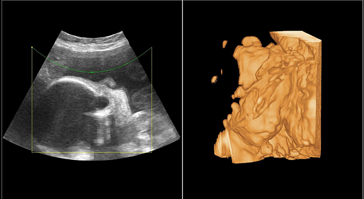
| Freehand 3D imaging | Support probe | convex probe, linear probe |
| Display model | :4 pictures | |
| Image Rotation X/Y/Z Axis | ||
| Multi-slice Visibility | ||
| Real-time 4D imaging | Support probe: 4D volume probe Adjustment of B mode parameters is switchable | |
| Gain | 0-100,Step 2 | |
| Display model | one image、two images、four images | |
| Image Rotation | X/Y/Z Axis Multi-slice Visibility Light&Shade inversion | |
| Smooth | 0-4, 4 level | |
| Threshold level | 0-129, Step 3 | |
| Transparency | 1-509,Step 10 | |
| Render type | 4 kinds,Surface、maximum、minimum、perspective | |
| Extended Imaging | Gain | 0-100,Step 2 |
| TGC | 8 segment adjustable | |
| Maximum focus point | ≥7, which can be moved throughout the whole process. | |
| Speckle reduction | 0-5,5 level | |
| Space Compound | 0-2,2 level(Linear probe: 3 level,don’t support cardiac probe) | |
| Dynamic range | 30-180,35 level,Step 5 | |
| Line density | Low、Middle、High,3 level | |
| Frame correlation | 0-4,4 level | |
| Noise reduction | 0-5,5 level | |
| Edge enhancement | 0-5,5 level | |
| Sound power | 2-10, 9 level | |
| Grey map | 0-67, 67 level | |
| False color | 0-67,67 level | |
| Image style | Soft-Comparison,2 level | |
| Extended level: | Maximum 72 leve | |
| Convex probe | 9 level | |
| Trans-vaginal probe | 72 level | |
| Micro-convex probe | 29 level | |
| Cardiac probe | 40 level | |
| 4D Volume probe | 17 level | |
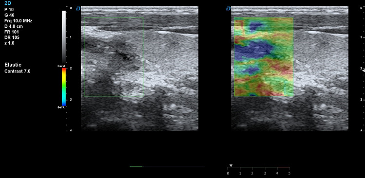
| Measurement and analysis function | General measurement | Distance, area, ellipse, cross line, angle, distance ratio, volume, Volume (ellipse), area ratio, diameter, joint angle |
| Cardiac | Automatic spectrum envelope、LV、Main Pulmonary artery diameter、RVEDd、RVEDs、LVM、LAV、HR、MVF、AO、AR、LVOT、TVF、Pulmonic valve、Pulmonary vein、RV、Doppler fetal heart sound、LVET、LVM、LVMI、AV | |
| Vascular | carotid intima (IMT), length stenosis ratio, area stenosis ratio, IMT (back wall), IMT (front wall) | |
| OB | Fetal routine、AFI、TW、GS、CRl、OFD、HL、ulna、NT、Fibula、Nbonel、Radial、Tibia | |
| GYN | uterus、cervix、corpus uteri/cervix uterus、left ovarian vein、right ovarian vein、dominant follicle、intima thickness | |
| Urologyseminal vesicle | :prostate、residual urine、left kidney、right kidney、left suprarenal vein、right suprarenal、left testis、right testis、left seminal vesicle、right | |
| liver、CHD、partal vein diameter、cholecyst、CBD、pancreas、spleen、Internal diameter of abdominal aorta、kidney Abdomen | ||
| Small parts | Thyroid | |
| Software package | Measurement package、Software package、Medical records management software package | |
| Graphic and text management system | Host build in 2 hard disk(SSD 120 1T),Start fast and stable | |
| Movie playback:≥1200 frames | ||
| Internal file information management system: can record patient number, name, check number, check date and so on, and can be searched and managed by numbering, checking number, name and so on. | ||
| Type of report is 16 | ||
| One key fast report graphic and text management | ||
| Interface | USB interface | 4 |
| HDMI interface | 1 | |
| RJ-45 interface | 1 | |
| Grounding wire interface | 1 | |
| DVD RW | 1 | |
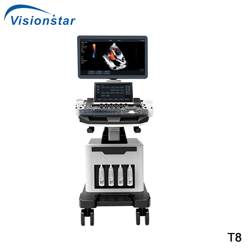
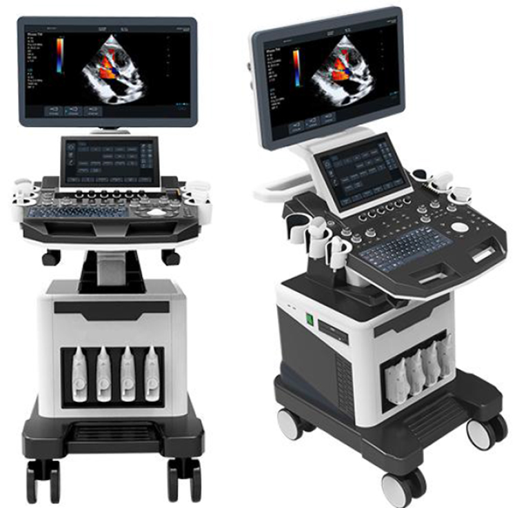
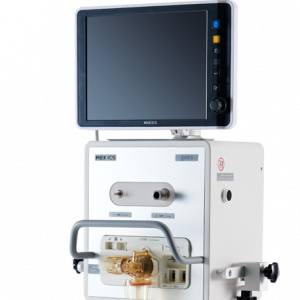
| Configuration | Trolley type full digital color Doppler ultrasound diagnostic system | |
| Probe: convex array probe (standard), linear probe (optional), Trans-vaginal probe (optional), cardiac probe (optional), 4D volume probe (optional) | ||
| ≥13 quick adjusting knobs | ||
Catalogue: PDF
See more products Vision Star Optical Co. ltd. brand.
Reviews
There are no reviews yet.