Specifications:
Clinical History
A 45-year old male presented with a lump in his left supraclavicular area. The swelling had been increasing in size over 6 months. Excision biopsy of the lump showed Hodgkin lymphoma (HL). Ten months later he was readmitted with left shoulder pain and swelling of his left arm. Examination revealed generalised lymphadenopathy with significant swelling in his left supraclavicular and axillary regions. He was treated with radiotherapy and Thiotepa chemotherapy. He developed vomiting. A subsequent barium meal showed duodenal obstruction from extrinsic lymph node compression. He continued to deteriorate and died 2 weeks after readmission.
Pathology
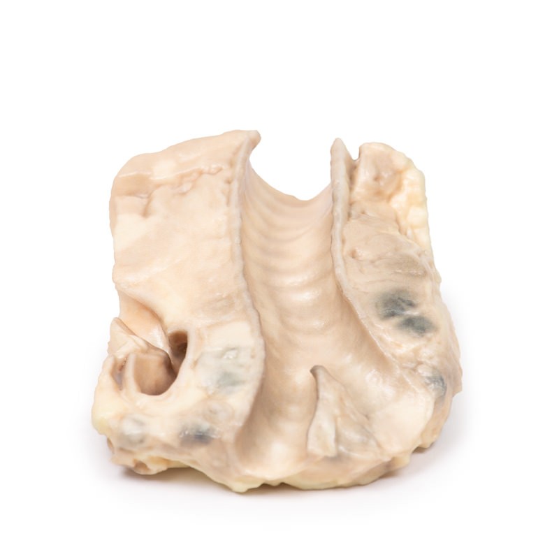
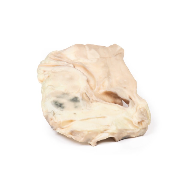
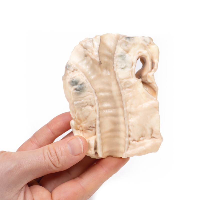
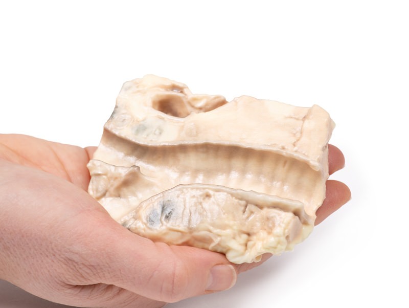
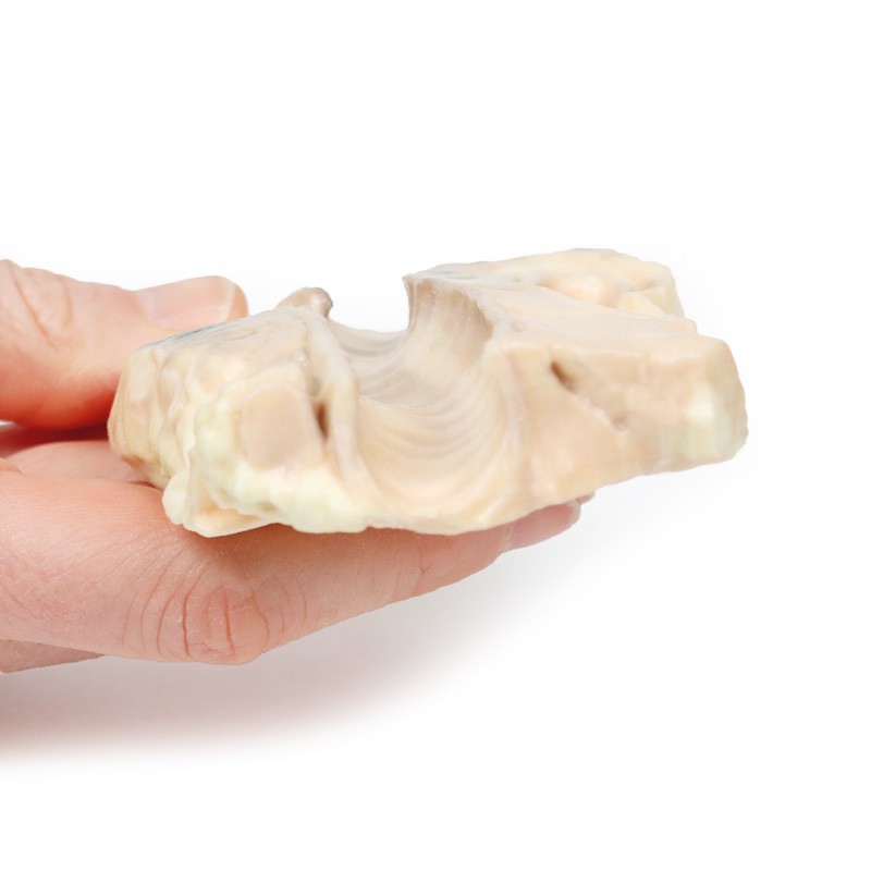
The 3D print shows the tracheal bifurcation with adjacent paratracheal and peri-bronchial lymph nodes. The trachea has been opened longitudinally and is viewed from behind. The para-tracheal lymph nodes are pale and matted (fused) together. Similar abnormal tissue is seen as a confluent pale mass on the left side of the trachea, above the aortic arch, which is seen cut in cross-section as a void space with branches arising. The peri-bronchial lymph nodes are also enlarged, and contain carbon pigment. The circumscribed small paler areas in the lymph nodes and extra-nodal tumour are foci of necrosis. There is an atheroma in the wall of the aorta but it is difficult to see in the 3D print.
Catalogue: PDF
See more products OpenMedis brand.
Reviews
There are no reviews yet.