Specifications:
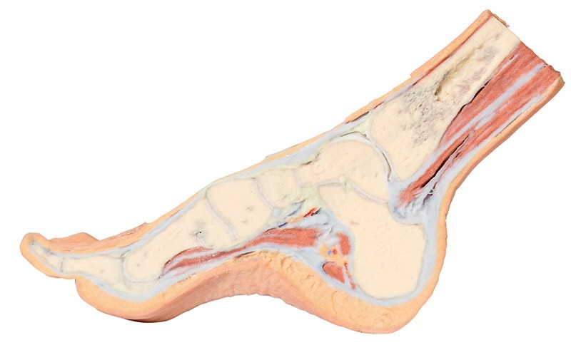
Product information: Foot – Parasagittal cross-section
This 3D printed specimen provides a parasagittal cross-section through the medial aspect of the right distal tibia and foot, displaying the skeletal structures of the medial longitudinal arch of the foot and surrounding soft-tissue structures. Proximally, the tendocalcaneus is visible superficial to the deep posterior compartment muscles and can be seen inserting into the posterior calcaneus. On the plantar surface of the medial arch, the plantar aponeurosis extends from the calcaneus towards the digits (were a sectioned lateral sesamoid is positioned at the head of the hallux). Part of the lateral head of the flexor hallucis brevis and muscular fibres at the origin of the flexor digitorum brevis and quadratus plantae are preserved (with the lateral plantar neurovascular bundle sectioned). Deep to these muscular portions is the flexor digitorum longus tendon (passing obliquely) near the calcaneus and neck of the talus, and the terminal insertion of the tibialis posterior tendon is visible at the articulation of the navicular and medial cuneiform.
Catalogue: PDF
See more products OpenMedis brand.

Reviews
There are no reviews yet.