Specifications:
Catalogue: PDF
See more products OpenMedis brand.
Features:
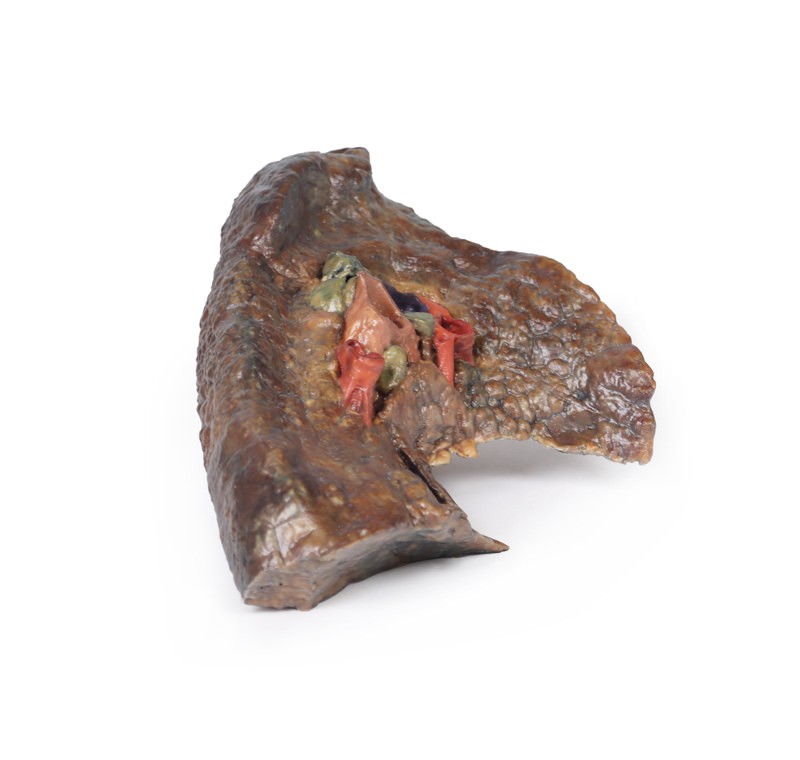
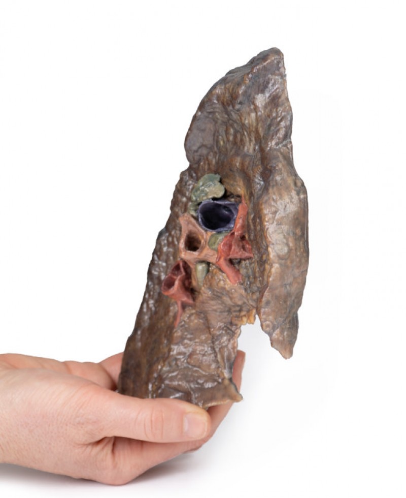
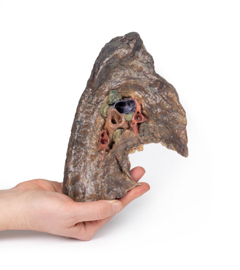
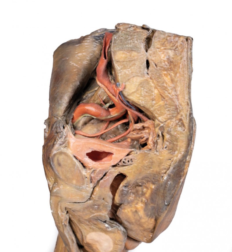
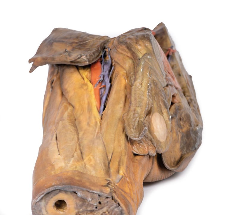
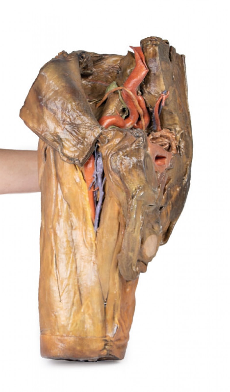
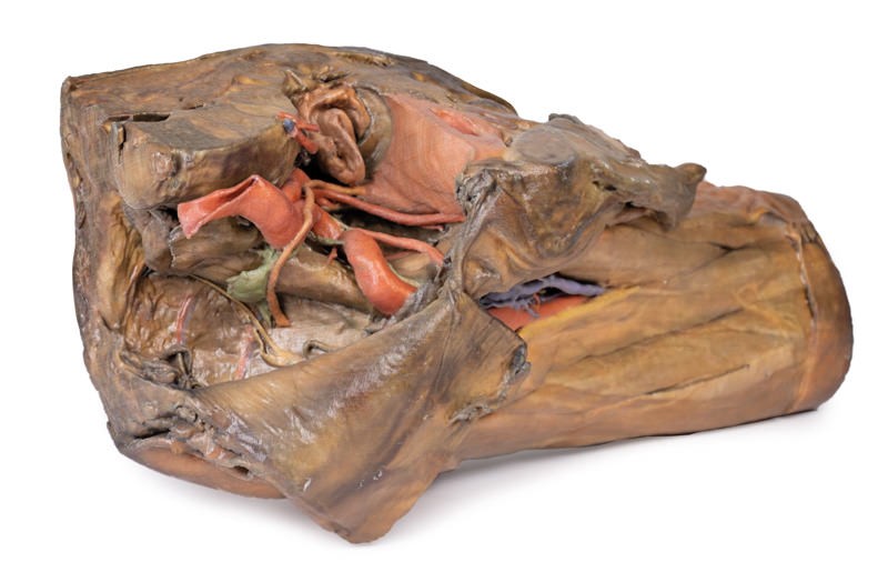
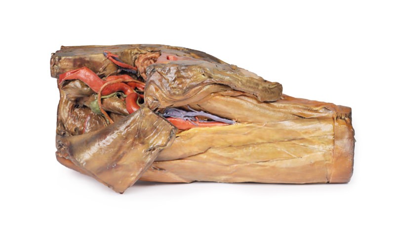
The hilum of a lung is the point at which visceral and parietal pleura meet and functions with the pulmonary ligament as the lungs only connection with the rest of the body. This connection includes the Pulmonary Artery, Superior and Inferior Pulmonary Veins, Main Bronchi, Nerves and Lymphatics.
As the definition of an artery involves carrying blood AWAY from the heart, this will be deoxygenated blood in the pulmonary system, in contrast with the systemic circulation. Similarly, veins carry blood TOWARDS the heart, meaning it will be oxygenated in the pulmonary system.
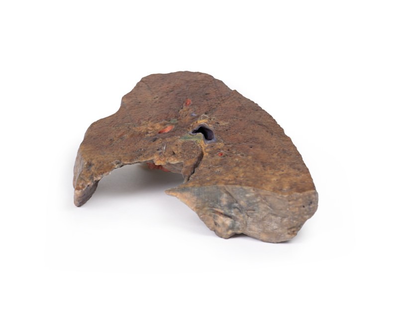
With the specimen cut in a sagittal plane in line with the cardiac notch, nerves are difficult to identify however, the impression from the arch of the aorta around the hilum can be seen alongside the left main bronchi and its subsequent divisions into lobar bronchi; found in this specimen more posterior in the hilum; the pulmonary artery and its divisions, located most superior; the superior and inferior pulmonary veins and their divisions which are most inferior and anterior in the specimen; the oblique fissure along the lateral surface of the specimen; various arteries, veins and bronchioles on the lateral surface; the diaphragmatic at the bottom and costal visceral on the posterior surfaces of the specimen and the pulmonary lymph nodes around the hilum on both the medial and lateral components of the lung.


Reviews
There are no reviews yet.