


Specifications:
Further Information
Tuberculosis (TB) is a chronic pulmonary and systemic infectious disease caused by Mycobacteria tuberculosis. Transmission most commonly occurs via inhalation of aerosolized droplets of M. tuberculosis. Risk factors for contracting TB include being an inhabitant of a developing country where the disease may be endemic, immunosuppression (e.g. HIV, steroid use, anti-TNF use and diabetes), chronic lung disease (e.g. silicosis), alcoholism and malnutrition.
After initial pulmonary infection of M. tuberculosis clinical manifestation varies. In 90% of individuals with an intact immune system they enter an asymptomatic latent infection phase. This latent TB may reactivate at any time in the patient‘s life. In the other 10% of patients, especially in the immunocompromised, they develop primary disease which is immediate active TB infection. Manifestations of primary TB include pulmonary infection symptoms (e.g. consolidation, effusion and hilar adenopathy) and extra pulmonary symptoms including lymphadenopathy, meningitis and disseminated miliary TB.
Secondary tuberculosis occurs when there is reactivation of previous latent TB infection. Around 10% of latent TB will reactivate usually during periods of weakened host immunity. Typical symptoms of reactivation are cough, haemoptysis, low grade fever, night sweats and weight loss.
The immune response against TB is mediated via TH1-cells stimulate alveolar macrophages to attack the mycobacteria. These macrophages surround the infection forming a ‘granuloma’ with central caseous necrosis.
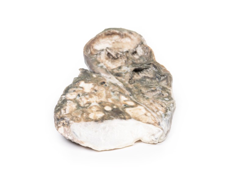
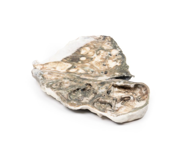
Secondary pulmonary TB may heal with fibrosis or progress as in this case. Progressive pulmonary TB sees erosion and expansion of the infectious lesion into adjacent lung parenchyma. This leads to evacuation of the caseous centre leading to fibrous cavitation. Erosion of blood vessels can occur causing haemoptysis. Post treatment of TB the tissue heals by fibrosis but does not recover the pulmonary architecture.
TB diagnosis is usually made with a clinical history and chest x-ray and multiple sputum cultures. Mantoux skin tuberculin test and serum interferon gamma release assay may also be used to help screen for infection. Biopsies may be taken of suspected infection site for culture to assist diagnosis. Treatment involves prolonged courses of multiple antibiotics, which depend on the antibiotic resistance of the infecting mycobacterium.
Catalogue: PDF
See more products OpenMedis brand.
Reviews
There are no reviews yet.