Specifications:
Product information: Foot – Deep plantar structures
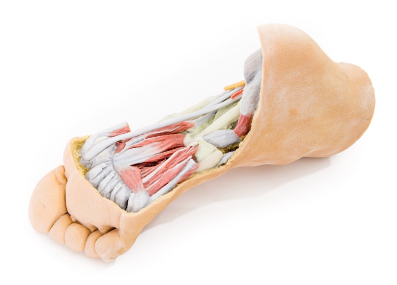
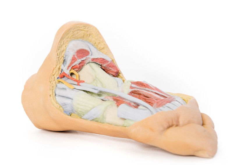
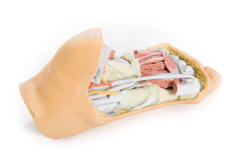
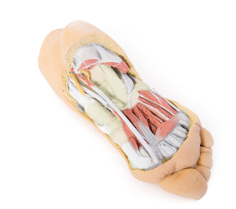
This 3D printed specimen provides a view of deep plantar structures of a right foot. Medially, the cut edge of the great saphenous vein is visible within the superficial fascia, just anterior to the cut edges of the medial and lateral plantar arteries and nerves overlying the insertion of the tibialis posterior muscle. The superficial fascias, the plantar aponeurosis, and superficial musculature have been removed to expose the deep (third layer) of musculature. The cut edges of the first, second and third layer muscles are preserved on the calcaneus for orientation, as is the cut tendon of the flexor digitorum longus muscle descending into the foot and the exposed distal tendons of the flexor digitorum longus and brevis muscles. The transverse and oblique heads of the adductor hallucis are visible deep to the tendon of the flexor hallucis longus muscle (surrounded by a complete lateral head and partial medial head of the flexor hallucis brevis muscle). The plantar interosseous muscles are visible deep to the adductor hallucis. Deep to the musculature the ligaments of the tarsal and metatarsal joint capsules are exposed, as well as the long and short plantar ligaments and the plantar calcaneonavicular ligament. On the lateral aspect, the abductor digiti minimi muscle has been sectioned to expose the insertions of the peroneus longus and brevis tendons are exposed.
Catalogue: PDF
See more products OpenMedis brand.


Reviews
There are no reviews yet.