Specifications:
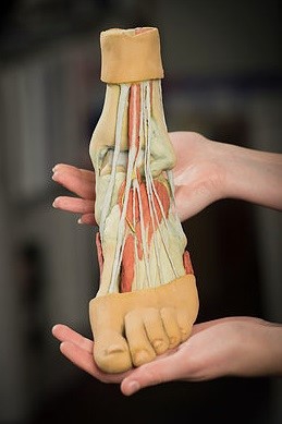
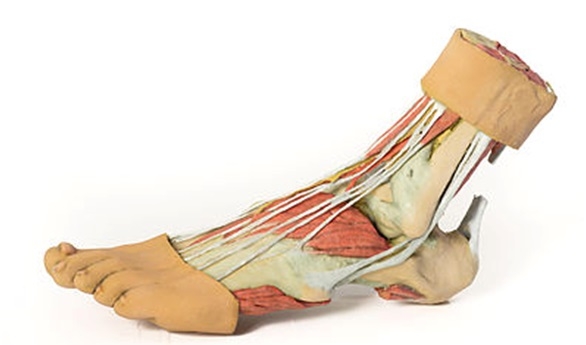
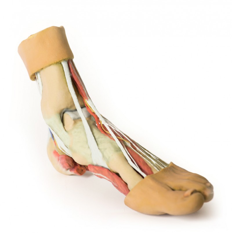
Product information: Foot – Structures of the plantar surface
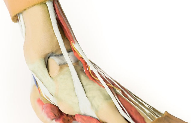
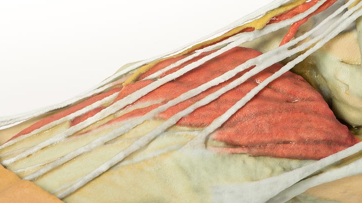
This 3D print records the anatomy of a right distal leg and the deep structures of the plantar surface of the foot. Proximally, the tibia, fibula, interosseous membrane, and leg muscles are discernable in cross-section. Medially, at the level of the ankle joint, the long tendons of the dorsi- and plantar-flexors are visible superficial to the capsular and extra capsular ligaments. The posterior tibial artery, veins, and tibial nerve are exposed through their course from the posterior leg to the plantar surface of the foot. Laterally, the course and insertion of the fibularis muscles (longus, brevis and tertius) are visible. On the dorsum of the foot, the anterior tibial artery and deep fibular nerve emerge from deep to the extensor hallucis longus just superficial to the extensor hallucis brevis and extensor digitorum brevis muscles. On the plantar surface of the foot, the plantar aponeurosis and portions of the superficial and deep musculature (flexor digitorum brevis, abductor hallucis, abductor digiti minimi, quadratus plantae) has removed between the calcaneous and bases of the metatarsals to display the course of the tibialis posterior, flexor digitorum longus, flexor hallucis longus, and fibularis longus tendons. The origins of both the flexor hallucis brevis and flexor digiti minimi brevis are visible, as are lumbribals arising from the flexor digitorum longus tendons.
Catalogue: PDF
See more products OpenMedis brand.


Reviews
There are no reviews yet.