


Specifications:
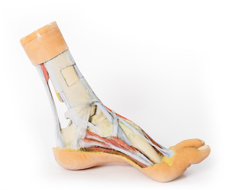
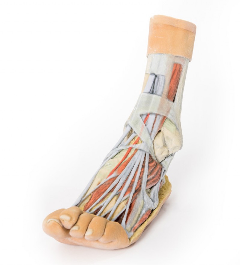
Product information: Foot – Superficial and deep dissection of the distal leg and foot
This 3D printed specimens preserves a mixed superficial and deep dissection of a left distal leg and foot. Posteriorly, the compartment muscles and neurovascular structures have been removed to isolate the tendocalcaneous and expose the body of the calcaneus. Medially, the tibialis posterior and flexor digitorum longus tendons are visible deep to the crural fascia, joined by the tendon of the flexor hallucis longus as the tendons passing deep to the flexor retinaculum (opened to demonstrate the tendon passage) to the medial foot. The adductor hallucis, medial head of the flexor hallucis brevis, and flexor digitorum brevis muscles are all exposed on the medial aspect of the foot. On the dorsum of the foot, both superior and inferior extensor retinacula are preserved, with the muscles of the anterior compartment of the leg extending to their distal attachments (including the fibularis tertius). The anterior tibial artery is exposed through to the dorsalis pedis artery. Deep to these long tendons are the extensor hallucis brevis and extensor digitorum brevis muscles, as well as the dorsal interosseous muscles. On the lateral aspect, both fibularis longus and brevis are visible deep to the crural fascia, with their tendons passing deep to both superior and inferior fibular retinacula. On the lateral margin of the foot the abductor digit minimi muscle is exposed.
Catalogue: PDF
See more products OpenMedis brand.


Reviews
There are no reviews yet.