Specifications:
Catalogue: PDF
See more products OpenMedis brand.


Features:
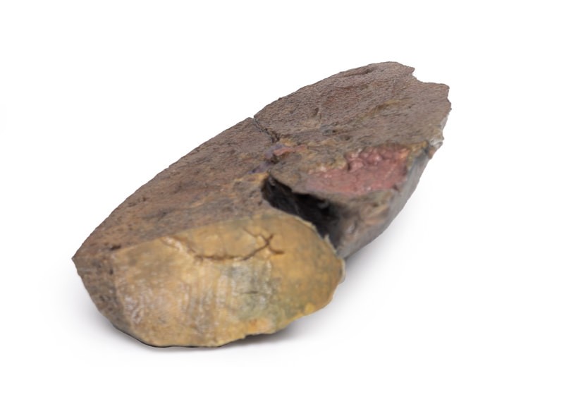
The lung has been dissected following a parasagittal plane, removing the mediastinal surface. Ordinarily, the pulmonary arteries, veins and bronchi can be observed entering the lung in the hilum – but the primary bronchi cannot be seen in this specimen as they have already divided substantially. It is unclear how far laterally the specimen has been dissected hence the bronchi subdivision level (secondary or tertiary) cannot be determined.
The cardiac impression is formed by the left ventricle of the heart resting on the mediastinal surface of the lung. Although the lung has been dissected following a parasagittal plane, the cardiac impression can still be observed as it is the most concave area of the medial surface of the lung.
The lung sits above the diaphragm, forming the concave diaphragmatic surface. The pleura has not been preserved in this specimen, but ordinarily, there exists a diaphragmatic recess bounded by the costal and diaphragmatic pleura. This would lie between the lung’s diaphragmatic impression and the diaphragm.


Reviews
There are no reviews yet.