Specifications:
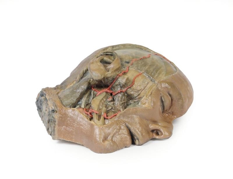
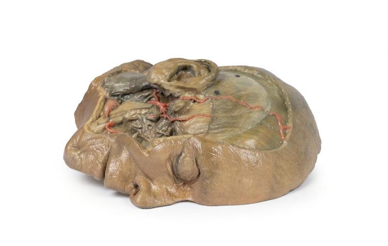
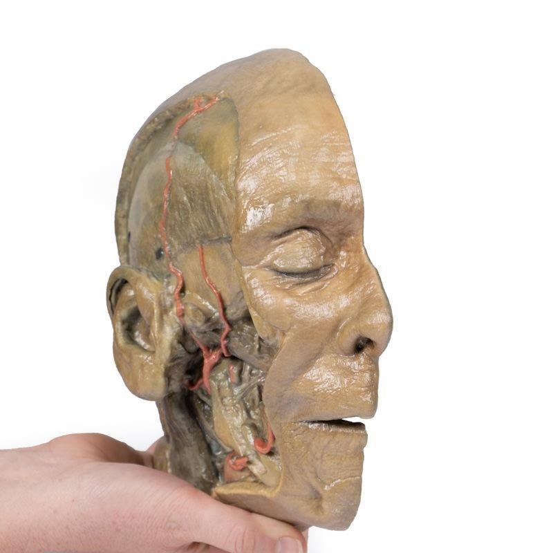
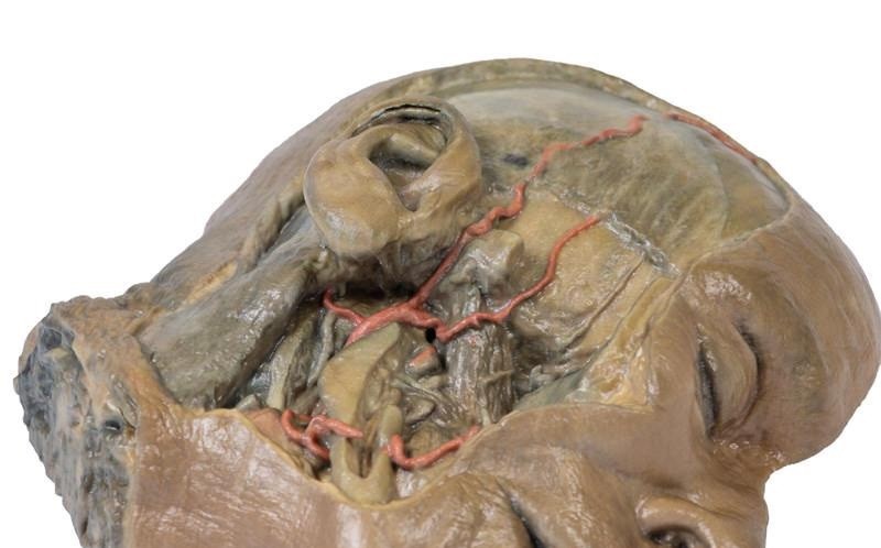
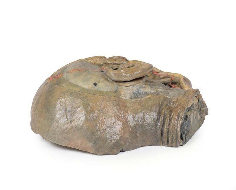
The deep level of dissection has exposed parts of the infratemporal fossa (through partial removal of the mandibular ramus and corpus) and dissection of retromandibular tissues. At the inferior margin of the dissection window, the cut edge of the retromandibular vein lies adjacent to the submandibular gland and the ascending path of the facial artery as it cross towards to angle of the mouth. Just superior to the cut retromandibular vein is the posterior belly of the digastric muscle, overlying a small exposure of the deeper internal jugular vein.
Just posterior to the retained ascending ramus of the mandible are the external carotid artery and the occipital artery (running in parallel prior to passing posteriorly). Tracing the external carotid artery superiorly, the posterior auricular artery, superficial temporal artery, and maxillary artery are all visible. The maxillary artery passes deep to the lateral pterygoid muscle and into the infratemporal fossa, reappearing superior to the lateral pterygoid as it passes into the pterygomaxillary fissure. Along its course, it gives rise to the posterior deep temporal artery, the inferior alveolar artery (which is exposed in the dissected mandibular corpus), the anterior deep temporal artery, and the posterior superior alveolar artery. Finally, the inferior alveolar nerve can be seen coursing within the opened mandibular corpus, and the lingual nerve resting on the medial pterygoid. The buccinator muscle is also retained, with the distal part of the parotid duct preserved as it enters the muscle towards the oral mucosa.
Catalogue: PDF
See more products OpenMedis brand.
Reviews
There are no reviews yet.